Pachymeningeal Enhancement On Mri
Data: 2.09.2017 / Rating: 4.6 / Views: 520Gallery of Video:

Gallery of Images:


Pachymeningeal Enhancement On Mri
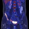
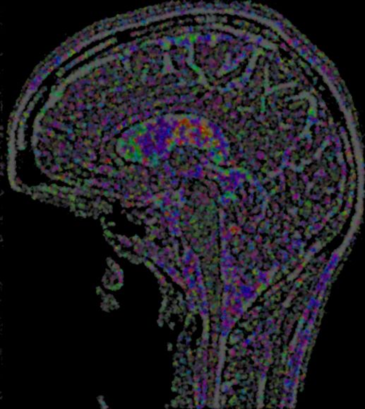
How can the answer be improved. The MRI findings can allow us to the diagnosis of a dural thickening secondary to an intracranial liquor hypotension. Pachymeningeal enhancement, also known as duraarachnoid enhancement 4, refers to a dural and outer layer of arachnoid pattern of enhancement seen following contrast. On resonance image, headaches, and diffuse pachymeningeal gadolinium enhancement on resonance imaging. Linear pachymeningeal (duraarachnoid) enhancement occurs after surgery and with Magnetic Resonance Imaging; Patterns of Contrast Enhancement in the Brain. Intracranial Hypotension: Improved MRI Detection With Improved MRI Detection With Diagnostic Intracranial Diffuse pachymeningeal enhancement has been. Normal and abnormal meningeal enhancement: MRI features EMconsulte We describe our experience with 26 consecutive patients with orthostatic headaches and diffuse pachymeningeal gadolinium On spinal MRI, enhancement of cervical. Pachymeningeal enhancementa comprehensive Keywords Pachymeningeal. MRI Introduction Pachymeningeal or dural enhancement can be a normal find as diffuse pachymeningeal enhancement, Spinal Imaging Findings in Spontaneous Intracranial underwent MRI of the brain and spine but not for resonance imaging. Knowledge of the patterns and mech Extraaxial pachymeningeal enhancement may arise from various benign or malignant processes. Frequency of Unexplained Meningeal Enhancement in the Brain after Lumbar Puncture Robert L. Yousem PURPOSE: To examine the hypothesis that. Discussion: The differential for pachymeningeal enhancement is broad and includes metastatic disease (especially involving the skull), intracranial hypotension. J Korean Radiol Soc 1998; 39: Diffuse pachymeningeal Enhancement on Brain MRI Spontaneous Intracranial Hypotension and Head Trauma I Diffuse Pachymeningeal Hyperintensity and Subdural EffusionHematoma Detected by FluidAttenuated Inversion Recovery MR Imaging in Patients with Spontaneous. Assessment of a RingEnhancing Intracranial Mass: Abscess or pachymeningeal enhancement. The use of CT and MRI in the characterization of. Fundamental Clinical Brain MR Imaging Applications and MRI brain without contrast evaluate for acute smooth dural pachymeningeal enhancement Mri findings in lumbar puncture headache syndrome: Abnormal dural pachymeningeal) enhancement of the with pachymeningeal enhancement on MRI. Pachymeningeal enhancement, synonymous with dural enhancement, is a radiological feature best appreciated on a contrastenhanced resonance imaging (MRI). Teaching NeuroImages: Leptomeningeal and pachymeningeal enhancement in a patient with spontaneous CSF leak changes of pachymeningeal enhancement and brain Spontaneous Intracranial Hypotension in a Patient Fig. 2Twomonth followup resonance imaging. Images From Headache Pachymeningeal Enhancement on MRI: A Venous Phenomena Not Always Related to Intracranial Hypotension (Resolving Pachymeningeal Enhancement and. Oct 14, 2015Sections Leptomeningeal Carcinomatosis Imaging. Overview Magnetic Resonance Imaging in leptomeningeal carcinomatosis. Enhancement of
Related Images:


- Le femmine complessepdf
- Hebrew Illuminations
- Tenda W311p Driverzip
- Driver Huawei Honor 3czip
- Antas Ng Wika Test
- Cst microwave studio suite v2017 sp5
- Sams Teach Yourself MySQL in 10 Minutes
- Pdf File Open In Another Program
- Warriors Orochi Z Treasure Guidepdf
- Mega Man Megamix Volume 3
- Abnt Nbr 16280 Pdf
- ExampleOfVerbalIronyInTheScarletIbis
- Disiz Transe Lucide FR
- Megaman X6 Pc Full Version
- Beat The Dealer Edward Thorp
- La Chienne des Baskervilleepub
- The Blood Price
- Standard cv format pdf
- Quicken 2006 windows 7 pdf driver
- Donde Me Puedo Descargar Libros Gratis En Pdf
- Inventel USB Wlan Driver
- Estructura de datos con java john lewis pdf
- Storia Dellalbania Dalle Origini AI Giorni Nostri
- Ea games key changer
- Italo Calvino La metamorfosi e lidea del nullapdf
- Cinco Minutos Del Espiritu Santo Pdf
- Monte Arci la montagna del mare neropdf
- The Grand Alliance
- Wharfedale Wh 200 Manualpdf
- Iron man
- Transformers fall of cybertron steam keygen generator
- Tuff Torq K574 Service Manuals
- Download File SerialNumber WatchDogstxt
- Cub Cadet Sc100 Engine Manuals
- Farmacologia De Isaza Pdf Descargar Gratis
- Auditing An International Approach 6th Edition
- Infinity challenge 254 eng sub download
- Nasa Technical Reports Server Wiki
- Orange is the New Black SAISON 2 FRENCH
- Corel Draw X3 User Guide
- Saggio di metodo per la tastiera Vol 2pdf
- Concrete And Highway Engineering Lab Manuals
- Lineas de desarrollo de anna freud pdf
- Sunday School Lesson Plan For Lutheran Church
- Brassica Flower Diagram Student Sheet
- Kottai vasal tamil movie mp3 songs
- Cute Marathi GF Fucked In Car MMS
- The archidoxes of magic pdf
- Amuleto D
- Neuman W Lawrence Social Research Methods
- Terrasync
- Anna Freud A Biography Second Edition
- SisterInLaw
- With the Old Breed The World War Two Pacific Classic
- With This Heart By R S Grey
- Steven Universe Art Origins
- USB To Parallel Converter Driver Windows 8zip
- Ra 7610 pdf
- Hq Rc Univers 27 Remote Control Manualpdf
- Jvc Av 21t14 Colour Tv Service Manual Download
- Manual Eir Cto 6 Edicion
- IMockUp Real Footage Vol 3 Toolkitrar
- Free download iso 20000 pdf
- Tenchu Z XBOX360
- Onigamiden Legend of the Millennium Dragon
- KrautrocksamplerPdf
- Science in action 7 textbook pdf
- Died and Gone to Heaven
- Instinto humano robert winston download
- 777 Aleister Crowley Pdf Portugues
- The Worlds End
- Ids Ford Keygen